Muscles Back (Upper Body)

MUSCLES OF THE HEAD (BACK)
Occipitalis

The occipital belly of epicranius muscle (also known as the occipitalis muscle) helps extend the scalp so that the eyebrows can come up and the forehead may wrinkle. This, along with the frontalis muscle is one of two parts, or bellies, of the epicranius. The epicranius is a musculofibrous layer that is wide and spreads across one entire side of the vertex of the skull, from the occipital bone to the eyebrow. The occipital belly is close to the occipital bone. This muscle takes on a thin, quadrilateral form and begins with tendinous fibres at the lateral two-thirds of the superior nuchal line of the occipital bone, and from the mastoid part of the temporal bone. The occipitalis ceases in the galea aponeurotica. The occipital belly exchanges information with the frontal belly by an intermediate tendon.
UPPER BODY MUSCLES
Trapezius

The trapezius is one of the major muscles of the back and is responsible for moving, rotating, and stabilizing the scapula (shoulder blade) and extending the head at the neck. It is a wide, flat, superficial muscle that covers most of the upper back and the posterior of the neck. Like most other muscles, there are two trapezius muscles – a left and a right trapezius – that are symmetrical and meet at the vertebral column.
The trapezius arises from ligaments at its origins along the nuchal crest of the occipital bone and the spinous processes of the cervical and thoracic vertebrae
It extends across the neck and back to insert via tendons on the clavicle, acromion, and spine of the scapula. The name trapezius is given to this muscle due to its roughly trapezoidal shape; the long base of the trapezoid is formed at the origins along the vertebrae and the short base forms at the insertions along the scapula and clavicle.
The trapezius can be divided into three bands of muscle fibres that have distinct structures and functions within the muscle.
- The superior fibres cover the posterior and lateral sides of the neck with their tendons connecting to origins along the occipital bone and insertions on the clavicle.
- Just below this region is the narrow band of the middle fibres, which extend from origins along the superior thoracic vertebrae and insert into the acromion process of the scapula.
- Finally, the inferior fibres cover a wide region of the back from their origins along the inferior thoracic vertebrae and insert into the spine of the scapula.
The functions of the trapezius are diverse and are best understood by actions of the individual bands of muscle fibres within the trapezius. The superior fibers typically act upon the scapula by elevating it (as in shrugging) or by bracing the shoulder when a weight is carried. When other muscles hold the scapula in place, the superior fibres of both trapezius muscles can extend the head at the neck by pulling the occipital bone closer to the scapula. The middle fibres work to retract and adduct the scapula by pulling the shoulder blade closer to the spine. The inferior fibres depress the scapula by pulling it closer to the inferior thoracic vertebrae. To rotate the scapula, the inferior and superior fibres work together to push the inferior angle of the scapula laterally and raise the acromion. Finally, the trapezius stabilizes the scapula to prevent extraneous movement by lightly contracting all of its fibre bands simultaneously.

The deltoid muscle is a rounded, triangular muscle located on the uppermost part of the arm and the top of the shoulder. It is named after the Greek letter delta, which is shaped like an equilateral triangle. The deltoid is attached by tendons to the skeleton at the clavicle (collarbone), scapula (shoulder blade), and humerus (upper arm bone). The deltoid is widest at the top of the shoulder and narrows to its apex as it travels down the arm. Contraction of the deltoid muscle results in a wide range of movement of the arm at the shoulder due to its location and the wide separation of its muscle fibres.
The deltoid has three origins: the lateral end of the clavicle, the acromion of the scapula at the top of the shoulder, and the spine of the scapula. Each origin gives rise to its own band of muscle fibers with the anterior band forming at the clavicle, the lateral fibers forming at the acromion, and the posterior fibres forming at the spine of the scapula. The bands merge together as they approach the insertion point on the deltoid tuberosity of the humerus.
The deltoid has three distinct functions that correspond to the three bands of muscle fibres. Contraction of the anterior fibres flexes and medially rotates the arm by pulling the humerus towards the clavicle. Flexion and medial rotation of the arm moves the arm anteriorly, as in reaching forward or throwing a ball underhand. The lateral fibres abduct the arm by pulling the humerus toward the acromion. Abduction of the arm results in the arm moving away from the body, as in reaching out to the side. Contraction of the posterior fibres extends and laterally rotates the arm by pulling the humerus toward the spine of the scapula. Extension and lateral rotation moves the arm posteriorly, as in reaching backwards or winding up to throw a ball underhand.
Infraspinitus

The infraspinatus muscle is one of the rotator cuff muscles. The stability of the shoulder joint is mainly provided by the tendons of the subscapularis, teres minor, infraspinatous, and supraspinatous muscles that together form the rotator cuff. The cuff is fused to the underlying joint capsule except inferiorly. Because of the lack of inferior stability, most dislocations or subluxations occur in this direction. The shoulder is most vulnerable when fully abducted and a force from a superior origin is applied.
Teres

The teres minor muscle rotates the arm laterally and assists in bringing it toward the body. As it draws the upper arm bone (humerus) up, it strengthens the shoulder joint. There are two teres muscles. The other one is the teres major muscle, a thick, flattened muscle that brings the arm toward the body and assists in extending it when the arm is in a flexed position. The teres major muscle also aids in rotating the arm but its function is just the opposite of the teres minor and other muscles in the rotator cuff.
The teres major muscle is a thick, flattened muscle that brings the arm toward the body and assists in extending it when the arm is in a flexed position. There are two teres muscles. The other one is the teres minor muscle, which rotates the arm laterally and assists in bringing it toward the body. As it draws the upper arm bone (humerus) up, it strengthens the shoulder joint. The teres major muscle also aids in rotating the arm but its function is just the opposite of the teres minor and other muscles in the rotator cuff.
Triceps
Triceps Brachii Lateral Head

The lateral head of the triceps brachii muscle is a muscle of the back of the arm, originating from the back of the humeral shaft and inserting at the elbow. The triceps brachii has three heads (connective immovable muscle) and is the only muscle on the back of the upper arm. It connects the humerus (upper arm bone) and the scapula (shoulder blade) to the ulna (longest of the forearm bones) and is the primary extensor of the elbow. The three heads are the lateral, the medial, and the long head.
Triceps Brachii Long Head

The long head of the triceps brachii muscle is a muscle of the back of the arm, originating from the scapula and shoulder to insert at the elbow. The triceps brachii has three heads (connective immovable muscle) and is the only muscle on the back of the upper arm. It connects the humerus (upper arm bone) and the scapula (shoulder blade) to the ulna (longest of the forearm bones) and is the primary extensor of the elbow. The three heads are the lateral, the medial, and the long head. The long head of the triceps brachii muscle, apart from the other triceps muscles, has a role in stabilizing the shoulder joint.
Extensor Muscles Of The Forearm
https://www.youtube.com/watch?v=7F4dHDwwvhQ
The extensor carpi radialis longus muscle runs along the lateral side of the forearm, connecting the humerus (upper arm bone) to the hand. It functions to extend the wrist and assists in abducting the hand.

Muscles Back (Upper Body)
Gluteus Maximus
The gluteus maximus muscle is located in the buttocks and is regarded as one of the strongest muscles in the human body. It is connected to the coccyx, or tailbone, as well as other surrounding bones. The gluteus maximus muscle is responsible for movement of the hip and thigh.
Standing up from a sitting position, climbing stairs, and staying in an erect position are all aided by the gluteus maximus.
Pain while rising to a standing position or lowering to sit may be caused by gluteus maximus syndrome. This syndrome is caused by a spasm in the muscle of the gluteus maximus. The pain usually disappears when sitting and affects only one side of the body.
Other causes of pain can be caused by an inflammation of the tendons or friction between the bones, tendons, and gluteus maximus muscle; these conditions are referred to as either bursitis or tendinitis. Treatments for these disorders include physical therapy or anti-inflammatory pills or injections. Physical therapists may try to put pressure on the joint of the gluteus maximus muscle and coccyx, or recommend exercises to reduce pain and improve range of motion.

Biceps Femoris

The biceps femoris is a double-headed muscle located on the back of thigh. It consists of two parts: the long head, attached to the ischium (the lower and back part of the hip bone), and the short head, which is attached to the femur bone.
The long head is a part of the hamstring muscle group that occupies the posterior section of the thigh. The hamstring muscles may be considered extensors of the thigh. The biceps femoris muscle is important for knee flexion, internal and external rotation, and hip extension.
Pain in the biceps femoris can be caused by several reasons. The most common condition is a strained muscle caused by improper lifting or too much exercise. Overuse of the biceps femoris can result in torn muscles and ligaments.
Adductor Magnus
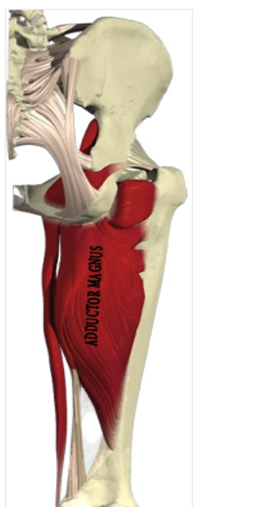
On the medial side (closest to the middle) of the thigh, the adductor magnus muscle creates the shape of a large triangle. As an adductor, it contracts and pulls the hip towards the body's midline. This action is a fundamental part of walking, sprinting, and a variety of other bipedal motions. The muscle also extends the hip. While an adductor, the muscle is often considered to be part of the hamstring group as well.
The muscle originates in the pelvic region; specifically, it arises from the pubis and the tuberosity of the ischium, which are also known as the sitz or sitting bones. Then, the muscle inserts into several parts of the femur bone.
Oxygenated blood arrives at the adductor magnus muscle via the obturator artery, which branches from the internal iliac artery. Once blood is depleted of oxygen, the obturator veins drain into the venal system.
For adductive motion, innervations come by way of the inferior branch of the obturator nerve. For hamstring functions, the muscle is served by the sciatic nerve.
Semitendonosis

The semitendinosus muscle is one of three hamstring muscles that are located at the back of the thigh. The other two are the semimembranosus muscle and the biceps femoris. The semitendinosus muscle lies between the other two. These three muscles work collectively to flex the knee and extend the hip.
The semitendinosus muscle begins at the inner surface of the base of the pelvis (known as the tuberosity of the ischium) and the sacrotuberous ligament. It inserts at the medial tibial condyle.
The semitendinosus muscle is comprised primarily of fast twitch muscle fibres. Fast twitch muscle fibres undergo rapid contractions for a short time period and easily wear themselves out.
The inferior gluteal artery and the perforating arteries bring oxygenated blood to the semitendinosus muscle. A segment of the sciatic nerve serves as the sensory and motor nerve for the muscle.
When the semitendinosus muscle becomes overly strained, a pulled hamstring results. There are three grades of pulled hamstring that are defined by how excessive the muscle tear is and the degree of pain and disability.
Gastrocnemius

The gastrocnemius muscle is a muscle located on the back portion of the lower leg, being one of the two major muscles that make up the calf. The other major calf muscle, the soleus muscle, is a flat muscle that lies underneath the gastrocnemius. Both the gastrocnemius and the soleus run the entire length of the lower leg, connecting behind the knee and at the heel. A third muscle, the plantaris muscle, extends two-to-four inches down from the knee and lies between the gastrocnemius and the soleus.
The gastrocnemius branches at the top behind the knee; the two branches are known as the medial and lateral heads. The flexing of this muscle during walking and bending of the knee creates traction on the femur, pulling it toward the tibia in the lower leg and causing the knee to bend. Both the gastrocnemius muscle and the soleus join onto the Achilles tendon, which is the strongest and thickest tendon in the human body. The tendon originates about six inches above the heel, running down the center of the leg to connect to the heel below the ankle.
Soleus

The soleus is the plantar flexor muscle of the ankle. It is capable of exerting powerful forces onto the ankle joint. It is located on the back of the lower leg and originates at the posterior (rear) aspect of the fibular head and the medial border of the tibial shaft.
The soleus muscle forms the Achilles tendon when it inserts into the gastrocnemius aponeurosis. The tibial nerves S1 and S2 innervate it; arterial sources include the sural, peronial, and posterior tibial arteries.
The soleus muscle is primarily used for pushing off the ground while walking. It may be exercised through calf raises while standing up or sitting down. The soleus is vital to everyday activities such as dancing, running, and walking. The soleus muscle helps to maintain posture by preventing the body from falling forward.
The soleus is also part of the skeletal-muscle pump, which is a collection of muscles that help the heart circulate blood. Veins within the muscles become compressed and decompressed as the muscles surrounding them contract and relax. This aids in venous return of blood to the heart.
WASLA Teacher Librarian of the Year- 2017: Jo-Anne Urquhart
- 2016: Lise Legg
WASLA Library Officer of the Year- 2012: Karen Notley